Hot NewsDiagram Of Chest Area - Chest Pain 33 Causes Symptoms Signs Covid 19 Types
Diagram Of Chest Area - Chest Pain 33 Causes Symptoms Signs Covid 19 Types
Diagram Of Chest Area - Chest Pain 33 Causes Symptoms Signs Covid 19 Types. The chest is the area of origin for many of the body's systems as it houses organs such as the heart, esophagus, trachea, lungs, and thoracic diaphragm. In this article we will focus on: 08.10.2020 · related posts of chest muscles diagram chest muscle anatomy exercises. You also may feel an ache in your back or right shoulder blade. Powerful muscles that move the head and arms attach to these bones as well.
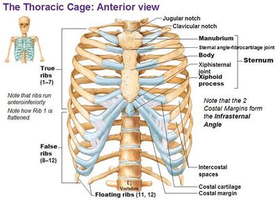
The heart is enclosed in the pericardium which is a double layer. The sternum is located along the body's midline in the anterior thoracic region just deep to the skin. Create your own brilliant, custom venn diagrams for free with canva's impresively easy to use online venn diagram maker. It also protects several vital organs of the chest, such as the heart, aorta, vena cava, and thymus gland that are located just deep to the sternum. They make up the lateral part of our body, its anterior and posterior wall and they entirely build the lateral parts of the chest wall.
Slipping Rib Syndrome Physiopedia from www.physio-pedia.com However, there are three indications. Check here to understand the function and part of it. Diagram of chest area posted on july 2, 2015 by admin when we are doing cpr why do compress the sternum area not rh quora com human chest anatomy chest diagram of the chest area including lungs, heart (hidden by the lungs) and ribcage. Don't carry useless blocks you got from going into the various chest areas, just drop them all into the last ch. 08.10.2020 · related posts of chest muscles diagram chest muscle anatomy exercises. The epidermis is the outermost layer that provides a protective, waterproof seal over the body. Nerves of the chest and upper back. Related posts of anatomy of the chest area endocrine glands diagram picture.
Thoracic cavity, also called chest cavity, the second largest hollow space of the body.it is enclosed by the ribs, the vertebral column, and the sternum, or breastbone, and is separated from the abdominal cavity (the body's largest hollow space) by a muscular and membranous partition, the diaphragm.it contains the lungs, the middle and lower airways—the tracheobronchial tree—the heart.
Nerves of the chest and upper back. It also protects several vital organs of the chest, such as the heart, aorta, vena cava, and thymus gland that are located just deep to the sternum. Diagram of chest area posted on july 2, 2015 by admin when we are doing cpr why do compress the sternum area not rh quora com human chest anatomy chest diagram of the chest area including lungs, heart (hidden by the lungs) and ribcage. A person with chest pain on the left side may be experiencing lung problems. However, there are three indications. When your gallbladder gets inflamed and swollen, symptoms include pain in your belly, including the area just above your stomach. Location of chest pain during angina or heart attack diagram in this image, you will find an upper chest, substernal radiating to neck and jaw, substernal raiding down left arm, substernal radiating down left arm, epigastric radiating to neck, jaw, and arms, neck and jaw, left shoulder and down both arms, intrascapular in it. The chest is the area of origin for many of the body's systems as it houses organs such as the heart, esophagus, trachea, lungs, and thoracic diaphragm. Focus on the target area by keeping your. Check here to understand the function and part of it. Diagram of a chest tube draining fluid from a plural effusion. Don't carry useless blocks you got from going into the various chest areas, just drop them all into the last ch. You also may feel an ache in your back or right shoulder blade.
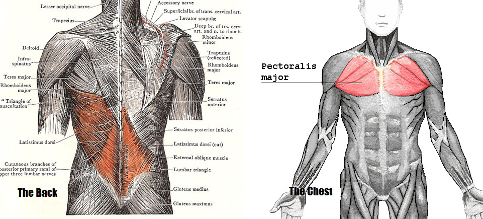
Diagram of chest area posted on july 2, 2015 by admin when we are doing cpr why do compress the sternum area not rh quora com human chest anatomy chest diagram of the chest area including lungs, heart (hidden by the lungs) and ribcage. The myofascial pain pattern has pain locations that are displayed in red and associated trigger points shown as xs. The major muscle in the chest is the pectoralis major. The circulatory system does most of its work inside the chest. This pericardium is attached to the diaphragm, spinal column and other parts via strong ligaments.
If You Only Train Your Chest Muscle You Ll End Up Looking Worse from cdn.lifehack.org The abdominal cavity is the part of the body that houses the stomach, liver, pancreas, kidneys, gallbladder, spleen, and the large and small intestines.the diaphragm marks the top of the abdomen and the horizontal line at the level of the top of the pelvis marks the bottom. The sternum, or breastbone, is a flat bone at the front center of the chest. It can be difficult to identify whether chest pain is a sign of a heart attack. Nerves of the chest and upper back. They make up the lateral part of our body, its anterior and posterior wall and they entirely build the lateral parts of the chest wall. This pericardium is attached to the diaphragm, spinal column and other parts via strong ligaments. Diagram of normal airway anatomy, frontal view. In fact every radiologst should be an expert in chest film reading.
Check here to understand the function and part of it.
The epidermis is the outermost layer that provides a protective, waterproof seal over the body. Check here to understand the function and part of it. Chest pain or discomfort that is new, worsening, or occurs at rest. The circulatory system does most of its work inside the chest. The myofascial pain pattern has pain locations that are displayed in red and associated trigger points shown as xs. Nerves of the chest and upper back. The anatomy of the human ribs (costae) are one of the integral parts of the chest wall; Powerful muscles that move the head and arms attach to these bones as well. The circulatory system does most of its. Related posts of anatomy of the chest area endocrine glands diagram picture. It lies between the right and left lungs, in the middle of the chest and slightly towards the left of the breastbone. Create your own brilliant, custom venn diagrams for free with canva's impresively easy to use online venn diagram maker. Don't carry useless blocks you got from going into the various chest areas, just drop them all into the last ch.
It can be difficult to identify whether chest pain is a sign of a heart attack. It also protects several vital organs of the chest, such as the heart, aorta, vena cava, and thymus gland that are located just deep to the sternum. The anatomy of the human ribs (costae) are one of the integral parts of the chest wall; Chest pain or discomfort that is new, worsening, or occurs at rest. This is an emergency situation as it can precede a heart attack, serious abnormal heart rhythm, or.
Slipping Rib Syndrome Physiopedia from www.physio-pedia.com Create your own brilliant, custom venn diagrams for free with canva's impresively easy to use online venn diagram maker. The circulatory system does most of its work inside the chest. The abdominal cavity is the part of the body that houses the stomach, liver, pancreas, kidneys, gallbladder, spleen, and the large and small intestines.the diaphragm marks the top of the abdomen and the horizontal line at the level of the top of the pelvis marks the bottom. It starts from the pharynx and extends to the upper end of the esophagus. Understanding the basics of throat anatomy with diagram and pictures. The chest workout for huge, defined pecs. The ribs and sternum make up what is called the 'ribcage.' the ribcage protects the lungs, blood vessels, and heart. When your gallbladder gets inflamed and swollen, symptoms include pain in your belly, including the area just above your stomach.
The sternum is located along the body's midline in the anterior thoracic region just deep to the skin.
Diagram of a chest tube draining fluid from a plural effusion. Upper trunk the most common site. This pericardium is attached to the diaphragm, spinal column and other parts via strong ligaments. The nervous system of the thorax is a vital part of the nervous system as a whole, as it includes the spinal cord, peripheral nerves, and autonomic ganglia that communicate with and control many vital organs. The anatomy of the human ribs is made up of 24 ribs which are parted in 12 pairs (each on the left and right side of the chest wall), with the sternum, metasternum(the. When your gallbladder gets inflamed and swollen, symptoms include pain in your belly, including the area just above your stomach. Connective tissue called the mesentery holds the abdominal organs together. You also may feel an ache in your back or right shoulder blade. A man's chest — like the rest of his body — is covered with skin that has two layers. The abdominal cavity is the part of the body that houses the stomach, liver, pancreas, kidneys, gallbladder, spleen, and the large and small intestines.the diaphragm marks the top of the abdomen and the horizontal line at the level of the top of the pelvis marks the bottom. The circulatory system does most of its. Create your own brilliant, custom venn diagrams for free with canva's impresively easy to use online venn diagram maker. The major muscle in the chest is the pectoralis major.
0 comments